[Previous Chapter] [Next Chapter]
[Table of Contents] [Home] [Glossary] [Video] [Help]
Chapter 18
INSTRUMENTATION:
STIMULATION AND MEASUREMENT OF ACCOMMODATION
Key words: Optometer, Stimascope, Scheiner
Outline
XII. Near Response: Pupil constriction, Accommodation and Vergence.
Part I: Accommodation (contí): Measurement of Accommodation
- Introduction
- Stimulus Optometers
Simple
Badal
Nagel
- Subjective Measures of Accommodation
Stigmascope
Scheiner pupil (Hardenger and Coincidence Optometers)
- Objective Measures of Accommodation
3rd Purkinje Image Brightness and Size
Retinal Contrast (Cannon R-1)
Scheiner Principal- Ophthalmatron and SRI
Retinoscopy
Introduction
Devices that control the focus of the retinal image for the purpose of measuring the power of the eye are called optometers. This is the root name for the profession of Optometry. Optometers are subjective if the patient judges the clarity of the retinal image and objective if a machine or the examiner does it. The same optical design can be used for objective or subjective measurement. Subjective optometers control the focus of the retinal image and are used to determine when a target is conjugate to the retina. Objective optometers measure the defocus or disconjugacy of the retinal image and the stimulus target. There are also measures of accommodation that measure characteristics of the crystalline lens such as front surface curvature via the third Purkinje image. There are several single lens stimulus optometers
Stimulus Optometers
Simple Optometer
The simple optometer is a plus lens placed in the anterior focal plane or spectacle plane of the eye. The virtual image of objects placed before the lens can be imaged from infinity to close to the spectacle plane, simply by moving the target from the anterior focal plane of the lens to the lens plane respectively. The virtual image distance is calculated from the Gaussian equation:
1/l + F = 1/lí where: l = object distance, lí = image distance, F = focal power
Whenever you measure accommodation amplitude by the push-up technique with a phoropter lens before one eye, you are using a simple optometer. The reciprocal of image distance to the optometer lens equals the accommodative stimulus in reference to the spectacle lens plane. One common technique that uses the phoropter as a stimulus optometer is retinoscopy. All of you will conduct retinoscopy through a working lens that is in the spectacle plane. You vary power of additional lenses until the retinal image is focused in the plane of your retinoscope and that is when the reflex appears to move infinitely fast. This is an application of a simple optometer where the working distance remains constant and the simple optometer lens power is varied. In the clinic, the chart remains at 6 m and lenses are varied with a trial frame or phoropter before the eyes to determine the ametropia. It would be possible to have a fixed lens and vary the distance of the chart in front of it. This is done in the simple optometer. For example, let a +10.00 D lens be placed in the spectacle plane before the eye. A chart at 10 cm would be at optical infinity and theoretically clear to an emmetrope. Moving the chart away from the lens would blur the letters. Moving the chart toward the lens would stimulate accommodation but the letters would remain clear between the far and near points. It goes without saying that values are in reference to spectacle plane refraction.
One problem with the simple optometer in the measurement of accommodation is that the image increases in size with proximity so that you have both size and blur cues to accommodation. If you want to eliminate size cues you can use one of two other optometers: the Badal Optometer or the Nagel Optometer.
Badal Optometer (Jules Badal, 1876, French Ophthalmologist)
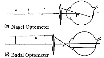
The Badal optometer utilizes a plus lens placed so that its posterior focal plane is coincident with the anterior focal plane of the eye. This instrument keeps image size constant while varying target distance and stimulus to accommodation. The optical system is telecentric in both the object and image space, that is, the rays are parallel.
 |
Fig 18.1 b) The Badal optometer has the focal plane coincident with the anterior focal plane of the eye. |
Nagel Optometer
The Nagel optometer is based on a similar concept. Here you have a plus lens whose posterior focal plane is coincident with the nodal point of the eye. It also keeps image size constant with changing object distance.
A variation of the Gaussian equation is needed to calculate accommodative stimulus with either the Badal or Nagal optometers because the Gaussian equation calculates the stimulus relative to the lens plane. The lens plane of these two instruments is not the spectacle plane of the eye or even the nodal point. To make the calculations relative to the eye you can use the Newton lens formula otherwise known as the lens makers equation. It is also used in lensometers. The formula allows us to define object and image distance relative to the anterior (x) and posterior (x') focal planes of the optometer lens and to specify the accommodative response or refractive error relative to a point coincident with the posterior focal point of the optometer lens. The formula states that x' * x'' = f' * f''. this can be reduced to 1/x''= x' * power^2. x'= f'-d where d is the distance of the target to the lens. By substitution, the accommodation stimulus equals 1/x''= (f'-d) * power 2. These equations and some examples have been worked out in the laboratory exercises.
Subjective Optometers
Stigmatoscopy
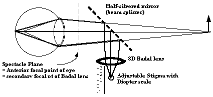
You can combine Simple and Badal lens optometers with various visual stimuli to enhance the sensitivity of subjective measures by improving sensitivity to blur detection. The stigmascope enhances blur perception with a small point source or stigma as the target viewed through the optometer lens. When the image of the point source is seen clearly and sharply, it is optically conjugate to the fovea. At the same time, this image may be introduced so that the eye can be fixating some other target which acts as the stimulus to accommodation such as Snellen chart. This instrument is called the stigmascope and is used in the labs on accommodation. A point source is seen superimposed with a beam splitter on an accommodative test target such as an acuity chart and the distance of the stigma is adjusted from the optometer lens until it is seen clearly while the subject maintains accommodation on the test chart. Bracketing the measures of positive and negative blur of the stigma allows you to estimate the accommodative response to the acuity chart stimulus with a precision of 0.1 D.
Fig 18.2 |
 |
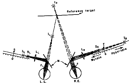
Fig 18.3 |
 |
Scheiner pupil
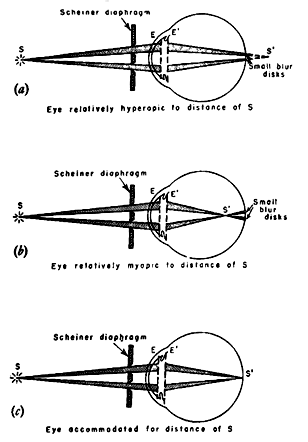
Christopher Scheiner was a Jesuit professor in Germany. He wrote the first formal treatise on the optics of the eye and so is sometimes called the father of physiological optics. Scheiner developed a double pupil that causes images viewed through it to appear double unless the image is in focus. Scheiner used this experiment, which you can perform with two pinholes in a card, to prove that the eye did accommodate rather than depend on universal focus for near vision. Furthermore it can be used to distinguish hyperopia from myopia.
 |
Fig 18.4 Scheiner pupil can be used to distinguish myopia from hyperopia. (See also Fig 16.6)
|
When the Scheiner pupil is combined with a Badal optometer it is called a coincidence optometer. With a coincidence optometer, the stigmascope is added to a two hole disc. When the point source is not in focus on the fovea, it will not only be blurred but will also be doubled. The greater the separation of the apertures in the Scheiner disc, the greater the separation of the images on the retina for a given nonfocal condition, thus increasing the precision. This is the principal behind a device called the Hardinger optometer. If a vertical prism is placed before one pupil you can assess accommodative errors using a vernier criterion. The Scheiner pupil was used by Thomas Young to evaluate his astigmatism. His astronomer friend Airy manufactured a cylindrical correction for his astigmatism. This principal is used in some of the objective infrared optometers and refractometers on today's market.
Objective Optometers
An objective optometer consists of two parts: an optical system to throw a bright image on the retina of the subject and an ophthalmoscope which, in being focused on the retinal image, discloses the state of refraction of the eye. Most autorefractors used clinically are objective optometers. Many use the Scheiner principal in which the two beams of the double Scheiner pupil are alternately interrupted (chopped) to facilitate the handling of the electronic signal mentioned below. The pair of infrared slit images on the retina are reflected out of the eye and are imaged on a twin plate photoelectric cell. The output signals oft he cell are amplified, filtered and after several other electronic steps are fed to a pen recorder. The record of accommodative response is written out with a noise level of about 0.05 D. it is designed to have a frequency response of 0 to 5 cps. This principal is used in research instruments such as the SRI objective optometer (Crane and Steele).
Other techniques use a servo mechanism to adjust the contrast of the retinal image until it reaches its peak. This is done in several meridians and then astigmatism can be calculated along with spherical refractive error. The Cannon R-1 system is based on this principal.
Another principal is to measure the size and brightness of the reflection of the front surface of the lens (3rd Purkinje image). This image becomes smaller and brighter with accommdation. It is used in laboratory measures (Kirchhof, 1950 ; Allen, 1949.; Stark-Young)
The best known method of objectively measuring the refractive state of the eye is retinoscopy or skiascopy. The skiascope can be used to measure accommodative response for a given stimulus. This is not done in the usual dynamic skiametry because the refractionist changes the stimulus as he changes lenses to obtain the desired reflex. In other words, he is increasing the optical stimulus distance until stimulus and response become approximately equal.
The most direct method to measure accommodative response is credited to Nott. In dynamic skiametry, one may seek neutrality by moving the skiascope back and forth instead of changing lenses. The position of the retinoscope at neutrality will yield the point conjugate to the retina.
Another important clinical measure is the Modified Estimate Method or MEM developed by Haynes. This technique is used to measure the lag of accommodation in children while they read. The child views a near point card that has a hole in its center. The refractionist conducts retinoscopy through the hole. If there is an accommodative lag, the reflex will appear hyperopic and there will be "with motion". As the patient views the letters on the near point card, the refractionist briefly places small amounts of plus lens before the eye and notes the plus value that yields a neutral reflex. The value of the lens that yields the neutral reflex equals the accommodative lag.
Review questions:
- What is the difference between subjective and objective measurements?
- What is a stigmascope?
- Describe the Scheiner pupil.
References:
- Fry & Reese (1941) The effect of fogging lenses on accommodation. Am. J. Opt. 18, 9-16.
- Fry (1945) The relation of pupil size to accommodation convergence. Am. J. Opt. monograph No. 11.
- Campbell & Robson (1959) High-speed infrared optometer. J. Opt. Soc. Am. 49(3), 268-272.
- Kirchhof (1950) A method for the objective measurement of accommodation speed of the human eye. Am. J. Opt. 27, 163-178.
- Allen (1949) An objective high speed photographic technique for simultaneously recording changes in accommodation and convergence. Am. J. Opt. 26, 279-289.
- Fry (1939) The Skiascopic determination of the limits of relative accommodation. Am. J. Opt. 13, 44-50.
- Fry (1940) Skiametric measurement of convergence accommodation. Optom. Weekly 31, 353-356.
[Previous Chapter] [Next Chapter]
[Table of Contents] [Home] [Glossary] [Video] [Help]