|
Chapter 5
ORBITAL GEOMETRY
ANATOMY OF EXTRAOCULAR MUSCLES Key Words: muscle planes, fields of action, primary, secondary and tertiary actions, agonist-antagonist pairs, Descartes-Sherrington law of reciprocal innervation.
Helpful references: Chapter 9 in Leigh and Zee / Chapter 5, section 1 in Adler
Outline
V. Orbital Geometry; Anatomy, Morphology, and Mechanics of EOM
- Semicircular canals: an ocular gyroscope
- Extraocular muscle orientation and actions
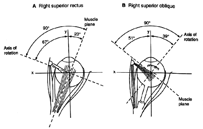
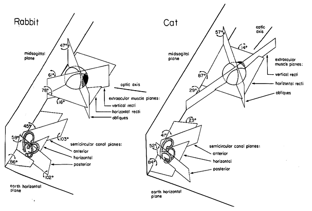
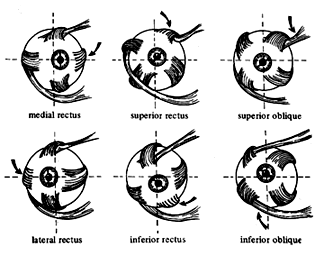
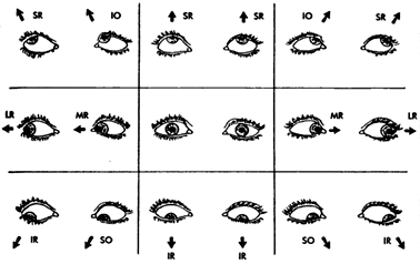
Points of insertion and angles with respect to globe (compare to cat & rabbit)
Actions- primary, secondary and tertiary
Conventional and soft pulley models
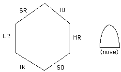
Pascal Benzene ring notation
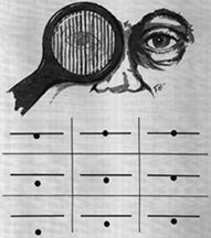
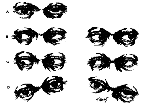
Diagnosis of noncomitancies - Hess screen, Lancaster test, Superior oblique palsies
- Laws of muscle action
Agonist and antagonist pairs- within eye interaction
Descartes-Sherrington law of reciprocal innervation
Force equilibrium between agonists and antagonists
Binocular yoking- between eye interaction
Hering's law of equal innervation
- (Outline continues in Chapter 6)
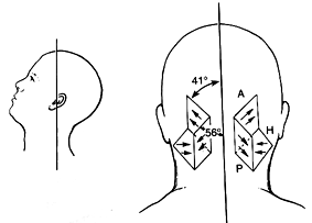
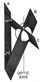
Semicircular canals: an ocular gyroscope
Comparative studies help us to understand the geometry of the orbit, globe and extraocular muscles. Afoveate eyes mainly served to survey the environment during locomotion. These eyes are visual gyroscopes that work exclusively with the vestibular canals and their function is to stabilize the image of the world as the animal moves about. It is helpful to look at the geometry of the vestibular canals to see this point. There are three pairs of canals (Horizontal, Anterior and Posterior). These canals lie in three plains that are nearly perpendicular to one another (i.e. orthogonal). This orthogonality improves their resolution of head motion in 3-D space. The horizontal canal lies in a plane nearly parallel to the ground. It is slightly elevated by 30 degrees. The anterior and posterior canals form a right angle with one another and they lie midway between the sagittal and frontal planes at an angle of 45 degrees. This orientation is consistent across the animal kingdom, even in fish, birds and reptiles. The stability of this orientation must have something to do with the mirror symmetry of organs in the head about the midsagittal plane. In addition, the orientation produces differential pairs on the two sides of the head so that when one canal is optimally stimulated its antagonist is inhibited. This opponence improves the linearity of the vestibular signal. |
|