[Previous Chapter] [Next Chapter]
[Table of Contents] [Home] [Glossary] [Video] [Help]
Chapter 16
ACCOMMODATION AND PRESBYOPIA
Key words: accommodation, pupil, lens, ciliary body, anterior, posterior, diopter
Note: An excellent review of the pupil is in Adler's Physiology of the Eye (Chapter 12). The following only addresses the accommodation and convergence components of the near response.
Outline
XII. Near Response: Pupil constriction, Accommodation and Vergence.
Part I: Accommodation
- Definition of Accommodation
- Comparative mechanisms of accommodation
Pupil size
Static variation in axial lenght (ramp retina)
Corneal shape and power
Position of lens altered by translation
Static shape of lens- anableps
Dynamic shape and power of lens
Index of refraction- isoindical surfaces of Gullstrand (n=1.403 & 1.386)
- Mechanisms in human
Relaxation Theory (dual mechanism) Helmholtz-Young-Fincham & Hess
capsular mechanisms from thickness variations
Constriction theory of Tscherning
- Anatomy of lens, capsule, zonules and ciliary body-isoindical layers
- Ocular changes during accommodation
Lens thickness increases .5 mm from 3.5-4mm
Lens diameter decreases .4 mm from10 to 9.6mm
Anterior radius of curvature decreases 5.5 mm from 11-5.5mm
Choroid pulls forward and displaces blind spot
Gravity influences lens position
- Innervation of Accommodation
- Amplitude and age (Donders data) (static response)
Amp= 18.5 - Age/3
Theories of presbyopia- Donders (changing myodiopter), Gullstrand (fixed myodiopter)
Mechanisms of presbyopia
Functional and Absolute presbyopia
Definition of Accommodation
Accommodation is the ability of the eye to vary its dioptric power in order to focus images at various distances on the retina. The variation in optical power makes the retina conjugate to a variety of target distances.
Mechanism of Accommodation
The mechanism of accommodation is one of the most varied in the animal kingdom. There are six different possible ways to vary optical power of the eye and all but one of these are used in nature.
- Vary axial length- statically with ramp retina and dynamically with accordion eye.
- Vary position of the lens (translation) within the eye.
- Vary corneal shape and power
- Vary lens shape and power
- Vary pupil size
- Vary index of refraction of the lens or cornea
Comparative Examples
1. The accordion eye is employed by the eel. It has a muscle that pulls back against the anterior sclera and shortens the eye moving the retina closer to the lens
2. The static eye or ramp eye is used by grazing animals like the horse. The top of the retina slopes back to accommodate near targets. Interestingly, ramp retinas can be developed in chickens by blurring part of the eye. It may result from an emmetropization mechanism in some animals.
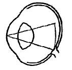 |
Fig 16.1 Near objects are focused on the superior retina, whereas far objects are focused on the inferior retina. |
3. Corneal shape and power can vary statically with high astigmatism. This is found with seals. It can also vary dynamically in some animals such as the owl.
4. Lens shape and position can be varied anterior posteriorly in most mammals including cats and raccoons. Primates vary accommodation by changing shape of lens with the ciliary body. Amphibious birds like the penguin, and mammal amphibians like the otter can also change lens shape by pinching the anterior lens surface with the iris. This process causes the largest changes (40-50D). This allows the animal to compensate for the large change in refractive index between air and water.
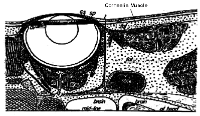 |
Fig 16.2 Cornealis muscle contracts, flattening cornea and pushing lens back toward the retina. (This is active negative accommodation.) |
 |
Fig 16.3 |
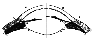 |
Fig 16.4 |
5. Some eyes also have an off-axis lens so that one optical path is more powerful than another in a dual optical system or physiological bifocal. The Anableps is a South American trout has this ability. It rests on top of water like a frog with eyes half submerged. The pupil viewing space above the water goes through a less powerful axis of the lens than light passing through the submerged pupil. The anableps can be seen at most large-city aquariums.
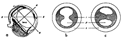 |
Fig 16.5 S= surface of water |
6. Pinhole pupils is another solution to accommodation, but it cuts down too much on light. The vertical slit pupil of the cat and reptiles is a solution for nocturnal animals.
7. The only mechanism not found in nature is changing refractive index.
History of Study in Humans
Accommodation was first demonstrated in man by Scheiner in 1619 using his renown double pupil apparatus. This system produces a single image of an external target when the retina is conjugate to the target, but it produces two images when the retina is not conjugate. In the figure below, a distant target (house) is seen as single in the unaccommodated eye (A) while the near target (pencil) is seen as double, and the reverse is true for the accommodated eye (B). This principal is used in many commercial automatic Optometers to measure refractive error objectively.
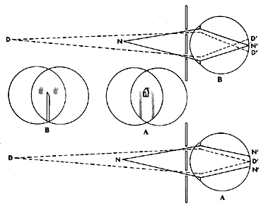 |
Fig 16.6 A) Unaccommodated |
Burow coined the term accommodation in 1841 referring to the process that changed the dioptric power of the crystalline lens to obtain and maintain clarity of an object of regard on the fovea.
Young was the first to demonstrate that changes in the crystalline lens were responsible for focus changes, although Descartes suggested this in 1677. Thomas Young ruled out the increased axial length and corneal shape changes as mechanisms in man. In 1849 Langenbeck showed the first measures of the Purkinje images to demonstrate changes in the front surface of the lens during accommodation.
At about this time, Helmholtz proposed his dual mechanism of accommodation that involved a passive mechanical lenticular mechanism during an active muscular contraction mechanism. This theory is referred to as the relaxation theory of accommodation as upheld by Fincham, Hess and Young. Helmholtz observed changes in the anterior chamber depth of up to 1.5 mm in young adults during accommodation. His model was supported by Young, Hess and Fincham. Fincham lived in recent times and developed ideas about the role of the lens capsule in accommodation.
The opponent theory is called the constriction theory by Tscherning who proposed the lens capsule was under increased force during accommodation. According to his theory, the Ciliary body was to pull the lens posteriorly against a rigid vitreous body and cause the front surface to bulge forward.
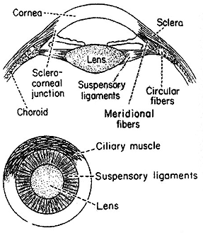 |
Fig 16.7 |
Anatomy
The lens is classified as one of the chambers of the eye. It is made up of alpha crystalline protein filaments that extend from sutures along the anterior to posterior pole. The nuclei of these filaments lie at the equator of the lens, and new fibers are laid down continuously throughout life. The older nuclear fibers become flattened with time so that the lens thickness only increases slowly with lens growth. These central compressed fibers exhibit a higher index of refraction than cortical fibers. This was shown by Gullstrand to be an important optical factor in accommodation. In his schematic eye he proposes a nuclear index of 1.406 and cortical index of 1.386. In fact there are many index zones, and these are referred to as isoindical surfaces. The variation of index of refraction acts as a powerful refractive mechanism. The power of the lens is much greater than computed by surface curvature and the average index of refraction of the lens. This is because light is continually refracted as it passes through the various layers of the lens. Astronomers used this principal by varying the density of air bubbles in their telescope lenses to increase its power. When we change surface curvature of the lens, actual power changes by more than would be predicted from the surface change alone. The lens layers are interlocked to produce both an elasticity and viscosity so that it tends to be sluggish and also to return to its original shape when forces are eliminated. When deformed or stretched it exhibits a spring like restoring force.
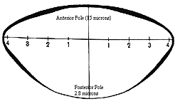 |
Fig 16.8 Notice the variation in thickness which helps round-out the lens during accommodation. |
The lens is surrounded by an elastic capsule made up of a collagen glycoprotein without an organized cellular structure. Capsule thickness varies with eccentricity. It is thinnest along the pole, and its posterior surface is thinner than anterior. In rest, the anterior is 15 microns and posterior pole is 2.8 microns. The periphery of the posterior side is 15.3 microns and the periphery of the anterior surface is 22 microns thick. (See Fig 16.8 above) The curvature is parabolic with the highest curvature in the center. This reduces peripheral aberrations (such as spherical and longitudinal chromatic aberrations). In a relaxed state the periphery is more powerful than the center and this produces positive spherical aberration. In the fully accommodated state the center is more powerful than the periphery and this produces negative spherical aberration. At about 3 D the center and periphery have equal power and spherical aberration is zero. At rest, the radius-of-curvature of the anterior surface is 12 mm and radius of curvature of the posterior surface is 5 mm. During accommodation the anterior surface increases curvature to match the 5 mm of the posterior surface. Fincham proposed these thickness and shape variations would help round the lens during accommodation.
Fig 16.9 |
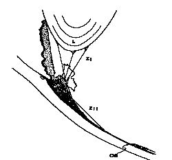 |
The lens is attached to the ciliary body and an elastic Bruch's membrane to the sclera via a suspensory ligament made up of a network of elastic cables called the Zonule of Zinn. The zonule has a fulcrum point where it attaches to the ciliary body. It extends beyond that point to attach to the sclera via elastic Bruch's membrane. When accommodation is relaxed, the anterior zonules are taut and place a stretching force on the capsule that applies a force normal to the surface of the lens, causing flattening. When the ciliary body contracts, the anterior zonules relax and relieve this stretching force.
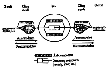 |
Fig 16.10 |
The ciliary muscle is a smooth muscle that has its origin at the scleral spur and it extends longitudinal fibers posteriorly to insert along the posterior zonule and Bruch's membrane. When the muscle contracts its longitudinal fibers bunch up anteriorly near the lens and there is also a contraction of radial fibers to provide a sphincter constriction that also reduces pull on the anterior zonules. The antagonist to this muscle is the mechanical elastic Bruch's membrane and the elastic span of posterior zonular fibers. When accommodation occurs the ciliary body must pull against the elastic restoring force of Bruch's membrane and the posterior zonules and stretch these structures. When accommodation relaxes, the restoring force of these mechanical structures elongates the ciliary body and allows the anterior fibers to be pulled back and stretch the lens to a flatter shape. The anterior and posterior zonule fibers meet at the ciliary processes at approximately a right angle. The posterior processes are running forward from Bruch's membrane toward the fulcrum point on the ciliary body and the anterior processes branch out from the fulcrum inward toward the middle of the eye. They branch into anterior, posterior and equatorial fibers that attach to the lens capsule in corresponding locations. When accommodation is minimal the span tents upward and pulls back on the anterior zonular fibers. When accommodation is stimulated, the ciliary body pulls the span forward and relieves the pull against the anterior fibers, allowing the lens to assume a more spherical shape. This sequence of events describes the relaxation theory of accommodation proposed by Helmholtz that has stood the test of time. There have been other theories such as a pulling back of the lens against the vitreous during accommodation causing the front surface to bulge forward (Tschermack theory) however Hess found no evidence of increased vitreal pressure during accommodation.
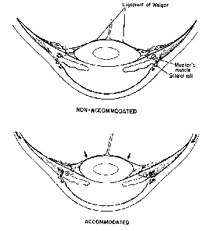 |
Fig 16.11 Changes in the eye during accommodation. |
Neural Innervation
There are two pathways of the autonomic nervous system that innervate accommodation. The dominant pathway is parasympathetic, cholinergic and uses acetylcholine at the synapses. The main motor nucleus is the Edinger Westphal that is located in the anterior portion of the III. Nerve axons project from here to the ciliary ganglion to form secondary axons that project to the ciliary muscle via the short posterior ciliary nerves. Some of these axons may not synapse in the CG and project directly from EW to ciliary muscle. There is also evidence of an inhibitory pathway controlled by the sympathetic branch of the autonomic nervous system. This system is a Beta adrenergeic class of synapse. Its pathway originates in the 8th cervical and first two thoracic regions of the spinal cord. Axons synapse at the superior cervical ganglion and project to the ciliary body via the long posterior ciliary nerves. This pathway appears to be responsible for relaxing accommodation below its resting state which is about 1.5 diopters. Thus, if a person sits in the dark, they exhibit a myopic shift in the far point that can be overcome with the negative accommodation stimulated by the sympathetic pathway.
 |
Fig 16.12 1) Optic nerve |
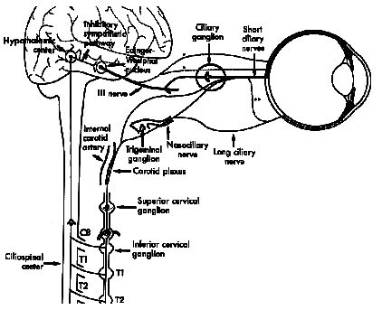 |
Fig 16.13 Sympathetic and parasympathetic (bold) pathways involved in accommodation. |
Review of what happens to the lens during accommodation
Four changes in the lens occur during accommodation:
- The lens thickens along the anterior-posterior axis by 3.5-4 mm
- The horizontal lens diameter decreases from 10mm to 9.6 mm
- Central anterior radius of curvature decreases from 11 to 5.5mm which causes the third Purkinje image to move forward and decreases in size.
- The choroid is pulled anteriorly causing displacement of the blind spot
Gravity effects that occur during accommodation:
- The anterior lens zonule slackens (slackening of lens capsule in aphake with capsule).
- The position of the lens is affected by gravity, it sinks .3mm (Hess and Fincham)
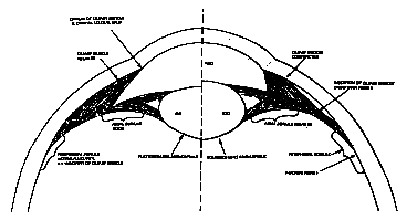 |
Fig 16.14 |
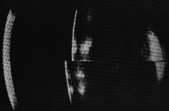 |
Fig 16.15 |
Amplitude and age
The continued growth of the lens produces a gradual reduction of amplitude of accommodation throughout life. The eye remains emmetropic during these changes and only its near point recedes. At birth we have about 18.5 diopters of accommodation and we lose 1 D every 3 years. This linear relationship was derived by Hofstetter using his and Donder's data for ages of 8 to 30 years. The regression equation describing this was calculated by Hoffstetter as: Amplitude = 18.5 - (age/3). This rule can be approximated by an easier to remember rule called the ìrule of 4'sî: Amplitude = 4X4 - (age/4).
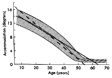 |
Fig 16.16 (Rule of 4ís shown by bold line, Hoffstetterís rule shown by dashed line.) |
Eventually the aging eye is unable to focus on near working distances. At this point, the eye is said to be presbyopic, but the definition depends on the patient's working distance. If the person has an outdoor occupation such as a forest ranger, presbyopia occurs later than if she or he works primarily with reading material at a desk. Clinically, it is defined in terms of a standardized working distance of 40 cm which corresponds to a stimulus to accommodation of 2.5 D. The amplitude of accommodation reaches 2.5 D at age 48 (on average). It reaches zero at age 55.5 (on average). However, most patients have trouble accommodating long before either age because of fatigue and excessive effort as the limit imposed by the near point is imposed. Generally, most people need a bifocal sometime in their early 40s. Subjective measures of amplitude of accommodation give larger values than objective measures because pupil constriction during subjective measures tends to keep the image from blurring. There can be discrepancies of over 1-2 diopters between objective and subjective measures. Subjective measures are made with good lighting and high contrast targets that your patient can see easily. However, when reading a low contrast map in a car at night, the patient may wish for bifocals.
What causes this reduction in accommodative amplitude and eventual progression to presbyopia? Any of a number of the sub-components of the accommodative process could be responsible. These include weakening of the ciliary body, loss of elasticity of Bruch's membrane, the zonules, the capsule, and even the lens. Growth of the lens takes up space in the eye and reduces the tautness of the lens zonules and finally, there is increased sclerosis or hardness of the lens which amounts to lost pliability. This increased growth is accompanied by an increased index of refraction and bigger changes in index as well as higher curvatures. In spite of this the degree of myopia doesn't increase with age. This is called the lens paradox. It may be explained by a more uniform overall index of refraction that causes most of the refraction to occur at the front and back surfaces of the lens rather than continuously at the many isoindical surfaces inside the lens.
The reduction of strength of the ciliary body is probably a secondary effect to loss of amplitude. We loose amplitude throughout life and it is unlikely that this is the result of muscle degeneration. However, if a muscle is not used, it atrophies. Loss of elasticity is a hypothetical possibility but it has not been tested. Increased diameter and thickness of the lens certainly reduces the efficiency of the zonules. Finally sclerosis is a factor but by itself it is not correlated with age to account for all of the effects.
Recently (ARVO 1997) it has been shown that the lens has reduced pliablity in presbyopia; however the main factor that reduces the amplitude of accommodation is the large diameter of the continually growing lens. It is very likely that surgical techniques will be developed in the next two decades that will restore accommodation in presbyopia with polymers injected into the empty lens capsule upon lens removal in cataract surgery. There have been attempts to sew scleral rings that expand the aperture that supports the lens and ciliary body. These have already been conducted on humans with some success. Several diopters of accommodation have been demonstrated following some of the surgical procedures, however residual refractive errors occur that require glasses, contact lenses, or refractive corneal surgery.
Review Questions:
- List 5 possible ways to vary the optical power of an eye.
- What is a Scheiner pupil?
- What are isoindical surfaces of the lens?
- What is the role of the Edinger Westphal nucleus in accommodation?
- Calculate the estimated amplitude of accommodation for a 30 year old.
[Previous Chapter] [Next Chapter]
[Table of Contents] [Home] [Glossary] [Video] [Help]