[Previous Chapter] [Next Chapter]
[Table of Contents] [Home] [Glossary] [Video] [Help]
Chapter 12
FIXATION EYE MOVEMENTS:
PHYSIOLOGIC NYSTAGMUS
Key Words: Physiological nystagmus, micro tremor, saccades, slow control drifts
Outline
- Fixation Eye Movements
Categories -- Micro tremors, Slow drifts, Micro saccades
Slow control mechanism
Fixation Eye Movements
If you observe the eyes during fixation, they appear motionless. However if you hold up a hand loop (plus lens) or use an eye movement monitor you will find the eyes are constantly moving. These movements are described with three categories: micro tremor, slow drifts, and fixation saccades (microsaccades).
The micro tremor is only a half minute of arc and cannot be seen with most eye movement monitors. The eyes oscillate at a high frequency of 50-60 Hz and the oscillations are independent in the two eyes. This disconjugate tremor is thought to result from the summed asynchronous activity of individual twitch fibers that make up extraocular muscle.
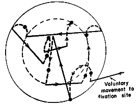
The other two components of physiological nystagmus are easily seen with most eye movement measures. The visual axis wanders about with slow drifts of approximately 6 arc min and a velocity of 1 arc min/sec, and then microsaccades rotate the eye back toward the target 3 times per second. Some of the drifts and micro saccades are error producing and some are error correcting. The error correcting nature is suggested by the increased frequency of microsaccades as the target drifts farther from the fovea. If you examine binocular steady fixation the drifts tend to correct for vergence misalignment of the two eyes and the microsaccades tend to correct monocular fixation errors. The micro saccades are yoked and the drifts can be either conjugate or disconjugate. Fixation saccades are also about 6 arc min in amplitude and they occur about 2-5 times each second. Their function is to correct monocular fixation errors that are produced by uncontrolled drifts or other saccades. The saccades of the two eyes are conjugate but can be slightly different in amplitude.
 |
Fig 12.1 Diameter of large circle is 10-min arc. Dashed lines are slow drifts, and solid lines are microsaccades. (Microsaccades attempt to correct postion errors caused by drift or other saccades.) |
It is not necessary to make saccades during fixation. People can be trained to control fixation solely with drifts but we make saccades to get the eye back on target as quickly as possible. This is a learned habit that comes with such tasks as reading and long-term visual inspection of fine detail. The saccades seen in the fixation trace are not necessary for steady fixation. Monkeys make fewer microsaccades than humans. A human subject can learn to make microsaccade if he/she is given feedback about their occurrence.
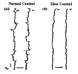
When the micro saccades do not occur, the eye still holds fixation using the slow drifts. This is referred to as a slow control mechanism. The slow control mechanism is likely to be under the control of the tonic integrators of the brainstem, and the lack of saccades is likely to result from the activity of fixation neurons at the rostral pole of the colliculus.
Fig 12.2 (Time on vertical axis) |
 |
Visual stimulation is not necessary for fixation but it helps. The eyes remain within 0.1 deg of the fixation mark in the light. In darkness, the same pattern of drifts and saccades occur, but the range increases by a factor of 20. The source of feedback used to control eye movements in darkness is proprioceptive from stretch receptors. (The feedback source was determined by severing the ophthalmic branch of the fifth nerve in the cat, resulting in large gross nystagmus in darkness but normal fixation in the light.) The control of fixation in darkness illustrates the corrective nature of both fixational drifts and saccades and that visual stimulation greatly improves fixation accuracy.
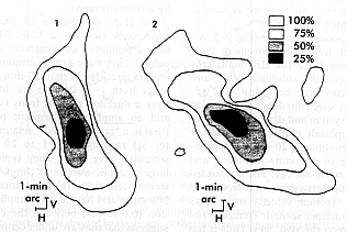
If the fixation nystagmus is large enough to see without the aid of an instrument it is probably a pathological condition such as latent nystagmus. The general rule is if you cannot see it with direct observation, it is probably normal. Otherwise, something is wrong.
Fig 12.3 Note the relatively small scale. (Normal fixation nystagmus is not easily observed.) |
 |
Review Question:
1. Describe the three categories of fixation saccades.
[Previous Chapter] [Next Chapter]
[Table of Contents] [Home] [Glossary] [Video] [Help]